1485
Views & Citations485
Likes & Shares
Recently,
WHO classification has proposed a uniform classification framework for all neuroendocrine
neoplasms (NENs). The classification is the distinction between
well-differentiated neuroendocrine tumor (NETs, previously called carcinoids)
and poorly differentiated neuroendocrine carcinomas (NECs). We describe
histopathological characteristics of a very rare case of the gallbladder
polypoid NET. The patient was a 49 year old man with upper abdominal pain.
Laparoscopic cholecystectomy was performed and the polypoid tumor was
pathologically diagnosed as NET, grade 1 (G1). The polypoid tumor was composed
of monotonous proliferation of tumor cells with weakly eosinophilic cytoplasm
and oval nuclei. Immunohistochemically, tumor cells were positive for
chromogranin A, synaptophysin, CD10 and MUC1. We speculated that the NET G1 had
arisen from enterochromaffin cells (also called Kulchitsky cells) because the
surrounding mucosa exhibited gastric-type metaplasia including enterochromaffin
cells.
Keywords: Neuroendocrine neoplasms, Neuroendocrine tumor, Neuroendocrine carcinomas, Polypoid, Gallbladder
INTRODUCTION
Recently, WHO has classified neuroendocrine neoplasms (NENs) into two
distinct groups: well-differentiated neuroendocrine tumor (NET) and poorly
differentiated neuroendocrine carcinoma (NEC) [1,2]. Primary NENs of
gallbladder are very rare. The majority of the previously reported gallbladder
cases were NECs with infiltrative/aggressive growth and poor patient’s
prognosis [3,4]. On the other hand, NETs have been reported as unusual cases.
Here, we describe histopathological characteristics of a gallbladder
polypoid tumor of the well differentiated NET.
CASE PRESENTATION
The patient was a 49 year- old Japanese man with upper abdominal pain,
and underwent laparoscopic cholecystectomy under the diagnosis of gallbladder
polyp. Macroscopically, the yellowish-white pedunculated polyp was located to
the gallbladder body, and measured 17 × 14 × 15 mm in size (Figure 1). Histologically, the polypoid tumor was composed of
monotonous proliferation of tumor cells with weakly eosinophilic cytoplasm and
oval nuclei (Figure 2). Nuclear
atypia is mild in a degree, and mitoses were few. In addition, the surface was
covered by the non-neoplastic gallbladder epithelium. The tumor cells are
limited within the mucosal polypoid lesion. There were no apparent foci of
lymphatic/venous invasion.
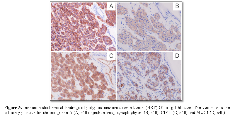
Immunohistochemically,
tumor cells were positive for chromogranin A, synaptophysin, CD10 and MUC1 (Figure 3); but negative for MUC5, AC, MUC6, MUC2 and p53 (Table 1). Ki-67 index is much less than 1%. The tumor cells
did not show any immunoreactivities for insulin, glucagon, somatostatin,
serotonin and gastrin. The surrounding non-neoplastic mucosa exhibited biliary
phenotype positive for CD10, as well as phenotype of gastric-type metaplasia
positive for MUC5AC and MUC6; but negative for intestinal phenotype (MUC2).
Based on the
above findings, the gallbladder tumor was diagnosed as NET, grade 1 (G1).
DISCUSSION
We describe a rare case of polypoid
NET G1 of gallbladder. Primary NETs of gallbladder are extremely rare,
estimated to account for 0.2% of all NETs [5,6]. NENs arise in most epithelial
organs with widely differing clinical features and histopathological findings
[2]. Previously, NENs were frequently called carcinoids including different clinicopathological
findings. The majority of the previously reported cases, called “carcinoids or
atypical carcinoids”, were diagnosed as NECs with infiltrative/aggressive
growth and poor patient’s prognosis [3,4].
Since 2010, WHO classifications have
proposed a uniform classification framework for all NENs. The key feature of
the classification is the distinction between well-differentiated NETs and
poorly differentiated NECs, while the term “carcinoid” is not recommended (Table 2). Therefore, many of the
recent reports have distinguished two histological types: NETs and NECs,
according to the WHO classification [3,4,7-9]. In the gallbladder cases, the
majority of the NEC cases are mixed neuroendocrine carcinomas; i.e., mixed tumors
of neuroendocrine carcinoma and non-neuroendocrine component. In the previous
reports, the non-neuroendocrine components were diagnosed as adenocarcinomas
(mixed adenocarcinoma-neuroendocrine carcinoma, MANEC). We have speculated the
NEC components arose through the phenotypical transformation of the
adenocarcinomas, because the adenocarcinoma components were frequently located
to the mucosal/superficial parts and the NEC components were usually seen in
the invasive/advanced areas.
In the gallbladder, the NET cases are fewer
than the NEC cases [2]. Therefore, histogenesis of gallbladder NET is still
poorly understood. Normal gallbladder mucosa lacks enterochromaffin cells, while
a few enterochromaffin cells are present in the mucous glands of the
gallbladder neck. On the other hand, gallbladder mucosa with chronic
cholecystitis frequently exhibits metaplastic changes, such as gastric-type
and/or intestinal-type metaplasia [11,12]. The metaplastic mucosa sporadically
has enterochromaffin cells (also called Kulchitsky cells, non-neoplastic
endocrine cells). In the present case, the surrounding gallbladder mucosa
exhibited gastric-type metaplasia positive for MUC5AC and MUC6. Therefore, we
speculated that anyone of the enterochromaffin cells (Kulchitsky cells) in the
metaplastic mucosa showed neoplastic changes and progressed to the NET G1.
In conclusion, we demonstrate a rare case of
polypoid NET G1 of gallbladder. Here, polypoid NET G1 should be added as one of
the differential diagnoses of gallbladder polyps.
ACKNOWLEDGEMENT
This study was supported by JSPS KAKENHI,
Grants-in-Aid from the Ministry of Education, Culture, Sports, Science and
Technology of Japan.
CONFLICT OF INTEREST
The authors declare
that they have no conflict of interest.
1.
Bosman FT, Carneiro F, Hruban RH, Theise ND (2010)
WHO classification of tumors of the digestive system. Lyon: IARC Press.
2.
WHO (2019) Digestive system tumor. WHO
classification of tumors, 5th Edn. Lyon: IARC Press.
3.
Fujii M, Saito H, Shiode J (2019) Rare case of a
gallbladder neuroendocrine carcinoma. Clin J Gastroenterol 12: 38-45.
4.
Skalický A, Vištejnová L, Dubová M, Malkus T,
Skalický T, et al. (2019) Mixed neuroendocrine-non-neuroendocrine carcinoma of
gallbladder: Case report. World J Surg Oncol 17: 55.
5.
Modlin IM, Lye KD, Kidd M (2003) A 5-decade
analysis of 13,715 carcinoid tumors. Cancer 97: 934-959.
6.
Albores-Saavedra J, Henson DE, Klimstra DS (2015)
Tumors of the gallbladder, extrahepatic bile ducts and Vaterian system. AFIP
Atlas of Tumor Pathology, series 4, fascicle 23. Washington DC: ARP Press.
7.
Koizumi M, Sata N, Kasahara N, Morishima K, Kaneda
Y, et al. (2011) Carcinoid tumor of the gallbladder: Report of two cases. Clin
J Gastroenterol 4: 323-330.
8.
Mezi S, Petrozza V, Schillaci O, La Torre V,
Cimadon B, et al. (2011) Neuroendocrine tumors of the gallbladder: A case
report and review of the literature. J Med Case Rep 5: 334.
9.
Chen H, Shen YY, Ni XZ (2014) Two cases of
neuroendocrine carcinoma of the gallbladder. World J Gastroenterol 20:
11916-11920.
10.
Mills (2019) Histology for pathologists. 5th Edn.
Philadelphia: Wolters Kluwer.
11.
Kijima H, Watanabe H, Iwafuchi M, Ishihara N (1989)
Histogenesis of gallbladder carcinoma from investigation of early carcinoma and
microcarcinoma. Acta Pathol Jpn 39: 235-244.
12.
Delaquerriere L, Tremblay G, Riopelle JL (1962)
Argentaffine cells in chronic cholecystitis. Arch Pathol 74: 142-151.
-
 Table 1
 -
Table 2


QUICK LINKS
- SUBMIT MANUSCRIPT
- RECOMMEND THE JOURNAL
-
SUBSCRIBE FOR ALERTS
RELATED JOURNALS
- Advance Research on Alzheimers and Parkinsons Disease
- Advance Research on Endocrinology and Metabolism (ISSN: 2689-8209)
- Journal of Pathology and Toxicology Research
- Journal of Psychiatry and Psychology Research (ISSN:2640-6136)
- Journal of Neurosurgery Imaging and Techniques (ISSN:2473-1943)
- Journal of Ageing and Restorative Medicine (ISSN:2637-7403)
- Journal of Rheumatology Research (ISSN:2641-6999)


